/ EN
Technical Support
| Contact Us
+1 312-802-6843
Technical Support
Contact Us
+1 312-802-6843
/ EN
Recover password
Professional Email
Verification Code
Send Code
Password
Recombinant Rabbit Monoclonal
Anti-HMGB2 Rabbit mAb
Catalog Number: PTM-6611
$ 285

Clone Number: JMMR-1856C

Host: Rabbit Clonality: Recombinant Monoclonal

Applications: WB IHC-P ICC/IF IP

Reactivity: Human, Mouse, Rat

Synonyms: High mobility group protein B2, High mobility group protein 2 (HMG-2), HMG2

Product Size
100 μl
Quantity

Shipping: Ambient temperature

Order online or send purchase order to info@ptmbio.com

FAQ Technical Support Protocols

General Information
Isotype IgG
Conjugate Unconjugated
Synonyms High mobility group protein B2, High mobility group protein 2 (HMG-2), HMG2
UniProt ID

P26583

Immunogen Synthetic peptide derived from human high mobility group protein B2
MW (kDa) 24
Specificity
Product Usage Information
Applications Dilution Recommended Species
WB 1:500 - 1:1000 Human, Rat
IHC-P 1:50 - 1:100 Human, Mouse, Rat
ICC/IF 1:50 Human
IP 1:25 - 1:50 Human
Properties
Purity Protein A purified
Constituents PBS, Glycerol, BSA
Storage Store at -20°C. Avoid freeze/thaw cycles.
Stability Stable for 12 months from date of receipt/reconstitution.
Target Information

Background

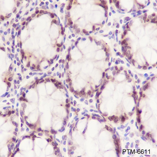
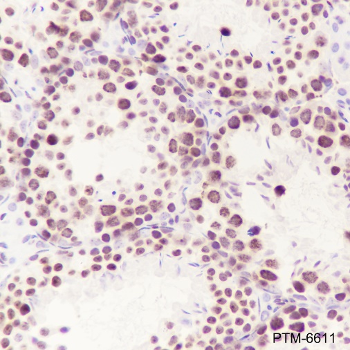
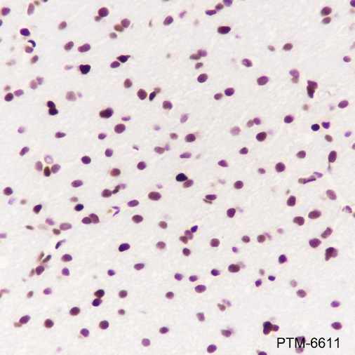
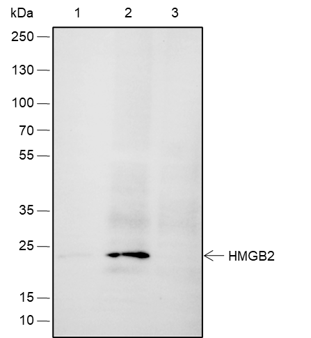
HMGB2 belongs to a family of highly conserved proteins that contain HMG box domains. HMGB2 is widely expressed during embryonic development, but it is restricted to lymphoid organs and testis in adult animals. HMGB2 facilitate the binding of Hox proteins, Oct proteins, p53, Rel proteins, and steroid hormone receptor proteins to their target gene promoters. Furthermore, HMGB2 interacts with RAG1 to facilitate RAG complex binding to the recombinant signal sequence (RSS) and stimulate DNA-bending and subsequent VDJ cleavage at antigen receptor genes. In addition to their functions in the nucleus, HMGB proteins play a significant role in extracellular signaling associated with inflammation. HMGB2 is secreted by myeloid cells and promotes proliferation and migration of endothelial cells by binding to the receptor for advanced glycation end products (RAGE).
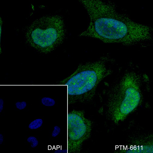
Cellular location

Cytoplasm, Nucleus, secreted

Images
WB

Blocking buffer: 5% NFDM/TBST
Primary Ab dilution: 1:1000
Primary Ab incubation: 2 hours at room temperature
Secondary Ab: Goat Anti-Rabbit IgG H&L pAb(HRP Conjugate)
Lysate: 1: HeLa, 2: HepG2, 3: PC-12
Protein loading quantity: 20 μg
Exposure time: 30 s
Predicted band size: 24 kDa
Observed band size: 24 kDa

IHC-P

Tissue: Human colon
Section type: Formalin-fixed & Paraffin - embedded section
Retrieval method: High temperature and high pressure
Retrieval buffer: Tris/EDTA buffer, pH 9.0
Primary Ab dilution: 1:100
Primary Ab incubation: 1 hour at room temperature
Secondary Ab: Anti-Rabbit and Mouse Polymer HRP (Ready to Use)
Counter stain: Hematoxylin (Blue)
Description: The brown color represents the positive signal observed with PTM-6611

Tissue: Mouse testis
Section type: Formalin-fixed & Paraffin - embedded section
Retrieval method: High temperature and high pressure
Retrieval buffer: Tris/EDTA buffer, pH 9.0
Primary Ab dilution: 1:100
Primary Ab incubation: 1 hour at room temperature
Secondary Ab: Anti-Rabbit and Mouse Polymer HRP (Ready to Use)
Counter stain: Hematoxylin (Blue)
Description: The brown color represents the positive signal observed with PTM-6611

Tissue: Rat cerebrum
Section type: Formalin-fixed & Paraffin - embedded section
Retrieval method: High temperature and high pressure
Retrieval buffer: Tris/EDTA buffer, pH 9.0
Primary Ab dilution: 1:100
Primary Ab incubation: 1 hour at room temperature
Secondary Ab: Anti-Rabbit and Mouse Polymer HRP (Ready to Use)
Counter stain: Hematoxylin (Blue)
Description: The brown color represents the positive signal observed with PTM-6611

ICC/IF

Cell line: HeLa
Fixative: 4% Paraformaldehyde
Permeabilization: 0.1% Triton X-100
Primary Ab dilution: 1:50
Primary Ab incubation: 4°C overnight
Secondary Ab: Goat Anti-Rabbit IgG
Nuclear counter stain: DAPI (Blue)
Description: The green color represents the positive signal observed with PTM-6611

IP

IP of HeLa cells extracts
IP Ab incubation: PTM6611, 4°C overnight, 1:50 dilution
WB Primary Ab incubation:
PTM6611, 1:1000 dilution, 2 hours at room temperature
Secondary Ab: Anti-Rabbit IgG for IP (HRP Conjugate)
Blocking buffer and concentration: 5%NFDM/TBST
Diluting buffer and concentration: 5%NFDM/TBST
1: 10% Input
2: IP with PTM6611
3: IP with Rabbit monoclonal IgG Isotype
Observed band size: 24 kDa
Exposure time: 60 s

Research Use

For research use only, not for use in diagnostic procedures.