/ EN
Technical Support
| Contact Us
+1 312-802-6843
Technical Support
Contact Us
+1 312-802-6843
/ EN
Recover password
Professional Email
Verification Code
Send Code
Password
Recombinant Rabbit Monoclonal
Anti-NeuN Rabbit mAb
Catalog Number: PTM-6825
$ 290

Clone Number: JRMR-10184-20B

Host: Rabbit Clonality: Recombinant Monoclonal

Applications: WB IHC-P mIHC-P

Reactivity: Human, Mouse, Rat

Synonyms: RNA binding protein fox-1 homolog 3, Fox-1 homolog C, Neuronal nuclei antigen (NeuN antigen), RBFOX3

Product Size
100 μl
Quantity

Shipping: Ambient temperature

Order online or send purchase order to info@ptmbio.com

FAQ Technical Support Protocols

General Information
Isotype IgG
Conjugate Unconjugated
Synonyms RNA binding protein fox-1 homolog 3, Fox-1 homolog C, Neuronal nuclei antigen (NeuN antigen), RBFOX3
UniProt ID

A6NFN3

Immunogen Synthetic peptide derived from human NeuN
MW (kDa) 46/48
Specificity Anti-NeuN Rabbit mAb recognizes endogeneous level of NeuN protein.
Product Usage Information
Applications Dilution Recommended Species
WB 1:500 - 1:1000 Mouse, Rat
IHC-P 1:500 - 1:1000 Human, Mouse, Rat
mIHC-P 1:500 - 1:1000 Human
Properties
Purity Protein A purified
Constituents PBS, Glycerol, BSA
Storage Store at -20°C. Avoid freeze/thaw cycles.
Stability Stable for 12 months from date of receipt/reconstitution.
Target Information

Background

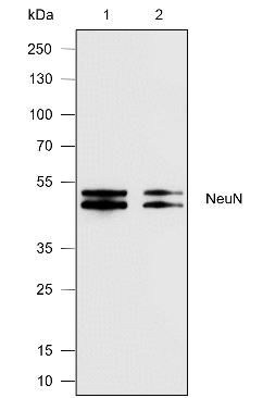
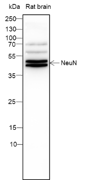
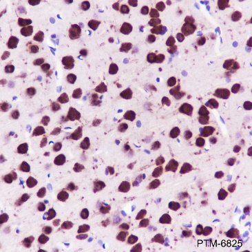
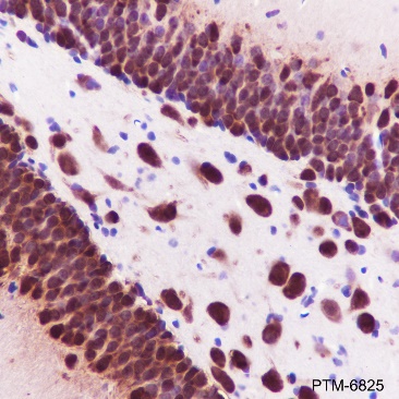
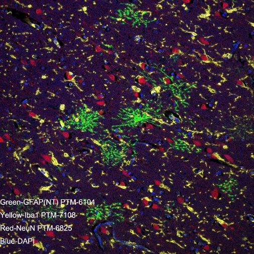
NeuN, also named as Fox-3 or RBFOX3, is a nuclear protein expressed in most post-mitotic neurons of the central and peripheral nervous systems. NeuN is not detected in Purkinje cells, sympathetic ganglion cells, Cajal-Retzius cells, INL retinal cells, inferior olivary, or dentate nucleus neurons. This neuronal protein was originally identified by immunoreactivity with a monoclonal antibody also called NeuN. Using MS-analysis, NeuN was later identified as the Fox-3 gene product. Fox-3 contains an RNA recognition motif and functions as a splicing regulator. Fox-3 regulates alternative splicing of NumB, promoting neuronal differentiation during development.
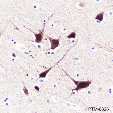
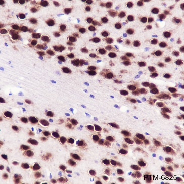
Cellular location

Nucleus, cytoplasm

Images
WB

Blocking buffer: 5% NFDM/TBST
Primary Ab dilution: 1:1000
Primary Ab incubation: 2 hours at room temperature
Secondary Ab: Goat Anti-Rabbit IgG H&L pAb(HRP Conjugate)
Lysate: 1: Mouse brain; 2: Rat brain
Protein loading quantity: 20 μg
Exposure time: 60 s
Predicted band size: 34 / 35 kDa
Observed band size: 46 / 48 kDa

IHC-P

Tissue: Human cerebral cortex
Section type: Formalin-fixed & Paraffinembedded section
Retrieval method: High temperature and high pressure
Retrieval buffer: Tris/EDTA buffer, pH 9.0
Primary Ab dilution: 1:1000
Primary Ab incubation: 1 hour at room temperature
Secondary Ab: Anti-Rabbit and Mouse Polymer HRP (Ready to Use)
Counter stain: Hematoxylin (Blue)
Description: The brown color represents the positive signal observed with PTM-6825

Tissue: Mouse cerebral cortex
Section type: Formalin-fixed & Paraffinembedded section
Retrieval method: High temperature and high pressure
Retrieval buffer: Tris/EDTA buffer, pH 9.0
Primary Ab dilution: 1:1000
Primary Ab incubation: 1 hour at room temperature
Secondary Ab: Anti-Rabbit and Mouse Polymer HRP (Ready to Use)
Counter stain: Hematoxylin (Blue)
Description: The brown color represents the positive signal observed with PTM-6825

Tissue: Mouse hippocampus
Section type: Formalin-fixed & Paraffinembedded section
Retrieval method: High temperature and high pressure
Retrieval buffer: Tris/EDTA buffer, pH 9.0
Primary Ab dilution: 1:1000
Primary Ab incubation: 1 hour at room temperature
Secondary Ab: Anti-Rabbit and Mouse Polymer HRP (Ready to Use)
Counter stain: Hematoxylin (Blue)
Description: The brown color represents the positive signal observed with PTM-6825

Tissue: Rat cerebral cortex
Section type: Formalin-fixed & Paraffinembedded section
Retrieval method: High temperature and high pressure
Retrieval buffer: Tris/EDTA buffer, pH 9.0
Primary Ab dilution: 1:1000
Primary Ab incubation: 1 hour at room temperature
Secondary Ab: Anti-Rabbit and Mouse Polymer HRP (Ready to Use)
Counter stain: Hematoxylin (Blue)
Description: The brown color represents the positive signal observed with PTM-6825

Tissue: Rat hippocampus
Section type: Formalin-fixed & Paraffinembedded section
Retrieval method: High temperature and high pressure
Retrieval buffer: Tris/EDTA buffer, pH 9.0
Primary Ab dilution: 1:1000
Primary Ab incubation: 1 hour at room temperature
Secondary Ab: Anti-Rabbit and Mouse
Polymer HRP (Ready to Use)
Counter stain: Hematoxylin (Blue)
Description: The brown color represents the positive signal observed with PTM-6825

mIHC

Tissue: Human cerebrum
Section type: Formalin fixed &
Paraffin -embedded section
Retrieval method: High temperature
and high pressure
Retrieval buffer: Tris/EDTA buffer,
pH 9.0.
Heat mediated antigen retrieval
(Tris/EDTA buffer, pH 9.0, 20
minutes) was used in between
rounds of tyramide signal
amplification to remove the antibody
from the previous round, to avoid any
cross-reactivity.
Primary ab dilution: PTM-6104
(1:1000), PTM-7108 (1:1000), PTM6825 (1:500)
Primary Ab incubation:
90 minutes at room temperature
Secondary ab: Anti-Rabbit and
Mouse Polymer HRP (Ready to use)
Nuclear counter stain: DAPI (Blue)
Comment: Color red is the positive
signal for NeuN (PTM-6825, TSA
570), color green is the positive
signal for GFAP(NT) ( PTM-6104,
TSA 520), color yellow is the positive
signal for lba1 ( PTM-7108, TSA
650). The section was incubated in
three rounds of staining; in the order
of PTM-6104,PTM-7108,PTM6825; each using a separate
fluorescent tyramide signal
amplification system.

Research Use

For research use only, not for use in diagnostic procedures.

Recommended Products
  • View More Images(5)
    $ 280
    Recombinant
    Anti-NeuN Mouse mAb
    Catalog #
    PTM-5313
    Host
    Mouse
    Clonality
    Recombinant Monoclonal
    Conjugate
    Unconjugated
    Reactivity
    Human, Mouse, Rat
    Applications:
    WB IHC-P ICC/IF FC IAP IP ChIP mIHC-P CUT&Tag