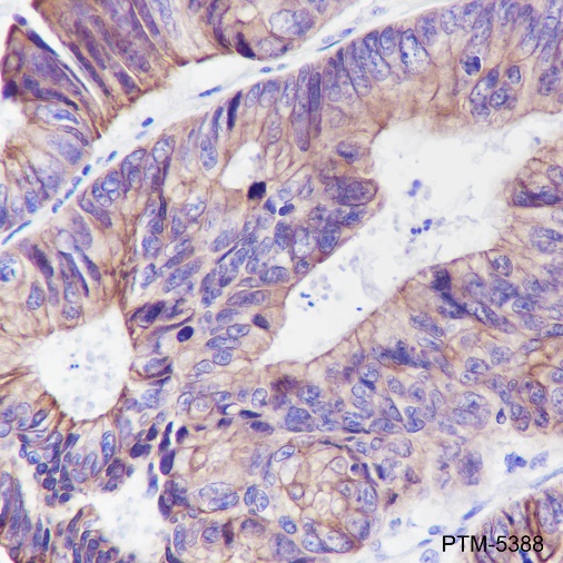
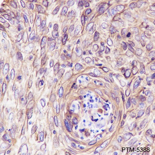
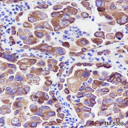
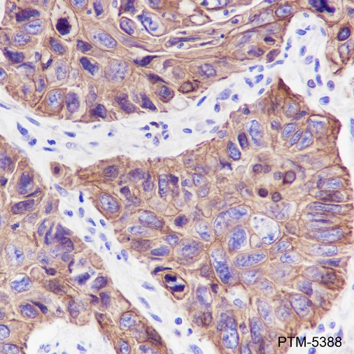
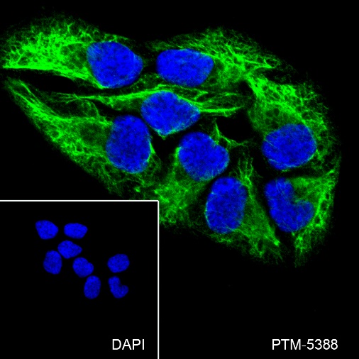
/ EN
Technical Support
| Contact Us
+1 312-802-6843
Technical Support
Contact Us
+1 312-802-6843
/ EN
Recover password
Professional Email
Verification Code
Send Code
Password
Recombinant Rabbit Monoclonal
Anti-Pan-Keratin Rabbit mAb
Catalog Number: PTM-5388
$ 290

Clone Number: Pan-CK (Cocktail)

Host: Rabbit Clonality: Recombinant Monoclonal

Applications: WB IHC-P ICC/IF mIHC-P

Reactivity: Human

Synonyms: Pan Cytokeratin

Product Size
100 μl
Quantity

Shipping: Ambient temperature

Order online or send purchase order to info@ptmbio.com

FAQ Technical Support Protocols

General Information
Isotype IgG
Conjugate Unconjugated
Synonyms Pan Cytokeratin
UniProt ID

/

Immunogen Recombinant human Pan-Keratin fragment
MW (kDa) 46-62
Specificity Anti-Pan-Keratin Rabbit mAb recognizes endogeneous level of Pan-Keratin protein.
Product Usage Information
Applications Dilution Recommended Species
WB 1:1000 Human
IHC-P 1:200 - 1:1000 Human
ICC/IF 1:50 Human
mIHC-P 1:200 - 1:1000 Human
Properties
Purity Protein A purified
Constituents PBS, Glycerol, BSA
Storage Store at -20°C. Avoid freeze/thaw cycles.
Stability Stable for 12 months from date of receipt/reconstitution.
Target Information

Background

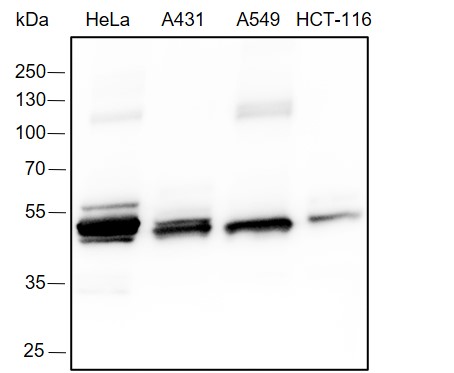
Keratins are a group of water-insoluble proteins (molecular weight range 40-70 K) that form 10-nm tonofilaments in a wide variety of epithelial cells. There are two types of cytoskeletal and microfibrillar keratin: I (acidic; 40-55 kDa) and II (neutral to basic; 56-70 kDa). The subunit composition of the keratin filaments varies with cell type, period of embryonic development, stage of histologic differentiation, cellular growth environment, and disease state.
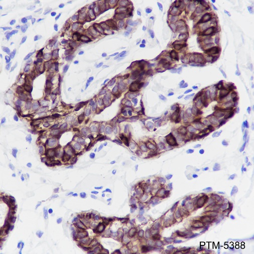
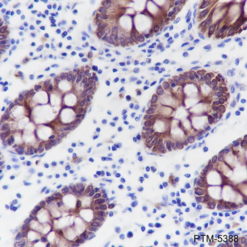
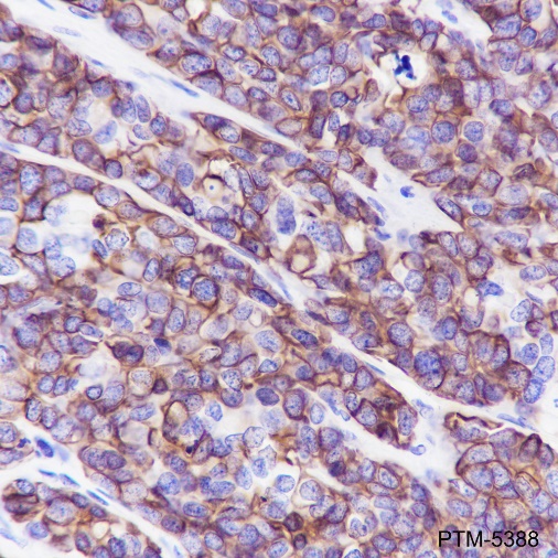
Cellular location

Membrane, Cytoplasm

Images
WB

Blocking buffer: 5% NFDM/TBST
Primary Ab dilution: 1:1000
Primary Ab incubation: 2 hours at room temperature
Lysate: HeLa, A431, A549, HCT-116
Protein loading quantity: 20 μg
Exposure time: 3 S
MW: 46-62 kDa

IHC-P

Tissue: Human breast
Section type: Formalin fixed & Paraffin -embedded section
Retrieval method: High temperature and high pressure
Retrieval buffer: Tris/EDTA buffer, pH 9.0
Primary Ab dilution: 1:1000
Primary Ab incubation: 1 hour at room temperature
Counter stain: Hematoxylin
Description: The brown color represents the positive signal observed with PTM-5388

Tissue: Human colon
Section type: Formalin fixed & Paraffin -embedded section
Retrieval method: High temperature and high pressure
Retrieval buffer: Tris/EDTA buffer, pH 9.0
Primary Ab dilution: 1:1000
Primary incubation condition: 1 hour at room temperature
Counter stain: Hematoxylin
Description: The brown color represents the positive signal observed with PTM-5388

Tissue: Human breast cancer
Section type: Formalin fixed & Paraffin -embedded section
Retrieval method: High temperature and high pressure
Retrieval buffer: Tris/EDTA buffer, pH 9.0
Primary Ab dilution: 1:1000
Primary incubation condition: 1 hour at room temperature
Counter stain: Hematoxylin
Description: The brown color represents the positive signal observed with PTM-5388

Tissue: Human colon cancer
Section type: Formalin fixed & Paraffin -embedded section
Retrieval method: High temperature and high pressure
Retrieval buffer: Tris/EDTA buffer, pH 9.0
Primary Ab dilution: 1:1000
Primary incubation condition: 1 hour at room temperature
Counter stain: Hematoxylin
Description: The brown color represents the positive signal observed with PTM-5388

Tissue: Human cervical carcinoma
Section type: Formalin fixed & Paraffin -embedded section
Retrieval method: High temperature and high pressure
Retrieval buffer: Tris/EDTA buffer, pH 9.0
Primary Ab dilution: 1:1000
Primary incubation condition: 1 hour at room temperature
Counter stain: Hematoxylin
Description: The brown color represents the positive signal observed with PTM-5388

Tissue: Human lung adenocarcinoma
Section type: Formalin fixed & Paraffin -embedded section
Retrieval method: High temperature and high pressure
Retrieval buffer: Tris/EDTA buffer, pH 9.0
Primary Ab dilution: 1:1000
Primary incubation condition: 1 hour at room temperature
Counter stain: Hematoxylin
Description: The brown color represents the positive signal observed with PTM-5388

Tissue: Human lung squamous carcinoma
Section type: Formalin fixed & Paraffin -embedded section
Retrieval method: High temperature and high pressure
Retrieval buffer: Tris/EDTA buffer, pH 9.0
Primary Ab dilution: 1:1000
Primary incubation condition: 1 hour at room temperature
Counter stain: Hematoxylin
Description: The brown color represents the positive signal observed with PTM-5388

ICC/IF

Cell line: HeLa
Fixative: 100% Ice-cold methanol
Permeabilization: 0.1% TritonX-100
Primary Ab dilution: 1:50
Primary incubation condition: 4°C overnight
Nuclear counter stain: DAPI (Blue)
Description: The green color represents the positive signal observed with PTM-5388

mIHC-P

Tissue: Human colon carcinoma
Section type: Formalin fixed & Paraffin-embedded section
Retrieval method: High temperature and high pressure
Retrieval buffer: Tris/EDTA buffer, pH 9.0. Heat mediated antigen retrieval (Tris/EDTAbuffer, pH 9.0, 20 minutes) was used in betweenrounds of tyramide signal amplification to remove the antibody from the previous round, toavoid any cross-reactivity.
Primary ab dilution: PTM-5388 (1:1000), PTM-5130 (1:500), PTM-5081 (1:1000)
Primary Ab incubation: 90 minutes at room temperature
Secondary ab: Anti-Rabbit and Mouse Polymer HRP (Ready to use)
Nuclear counter stain: DAPI (Blue)
Comment: Color red is the positive signal for Pan-Keratin ( PTM-5388, TSA 570), color greenis the positive signal for CD68 ( PTM-5130, TSA 520), color yellow is the positive signal for CD8 alpha ( PTM-5081, TSA 650). The sectionwas incubated in three rounds of staining; intheorder of PTM-5388,PTM-5130,PTM-5081; each using a separate fluorescent tyramidesignal amplification system.

Tissue: Human breast carcinoma
Section type: Formalin fixed & Paraffin-embedded section
Retrieval method: High temperature and high pressure
Retrieval buffer: Tris/EDTA buffer, pH 9.0. Heat mediated antigen retrieval (Tris/EDTA buffer, pH 9.0, 20 minutes) was used in between rounds of tyramide signal amplification to remove the antibody from the previous round, to avoid any cross-reactivity.
Primary ab dilution: PTM-5388 (1:1000), PTM-5130 (1:500), PTM-5081 (1:1000)
Primary Ab incubation: 90 minutes at room temperature
Secondary ab: Anti-Rabbit and Mouse Polymer HRP (Ready to use)
Nuclear counter stain: DAPI (Blue)
Comment: Color red is the positive signal for Pan-Keratin ( PTM-5388, TSA 570), color green is the positive signal for CD68( PTM-5130, TSA 520), color yellow is the positive signal for CD8 alpha ( PTM-5081, TSA 650). The section was incubated in three rounds of staining; in the order of PTM-5388,PTM-5130,PTM-5081; each
using a separate fluorescent tyramide signal amplification system.

Research Use

For research use only, not for use in diagnostic procedures.