/ EN
Technical Support
| Contact Us
+1 312-802-6843
Technical Support
Contact Us
+1 312-802-6843
/ EN
Recover password
Professional Email
Verification Code
Send Code
Password
Recombinant Rabbit Monoclonal
Anti-Crotonyl-Histone H4 (Lys8) Rabbit mAb
Catalog Number: PTM-522RM
$ 420

Clone Number: 36H1L2

Host: Rabbit Clonality: Recombinant Monoclonal

Applications: WB IHC-P ICC/IF ChIP

Reactivity: Human, Mouse, Rat

Synonyms: H4K8cr

Product Size
100 μl
Quantity

Shipping: Ambient temperature

Order online or send purchase order to info@ptmbio.com

FAQ Technical Support Protocols

General Information
Isotype IgG
Conjugate Unconjugated
Synonyms H4K8cr
UniProt ID

P62805

Immunogen
MW (kDa) 11
Specificity Anti-Crotonyl-Histone H4 (Lys8) Rabbit mAb detects histone H4 only when it is Crotonylated at Lys8.
Product Usage Information
Applications Dilution Recommended Species
WB 1:500 - 1:2000 Human, Mouse, Rat
IHC-P 1:100 - 1:500 Human, Mouse, Rat
ICC/IF 1:50 Human
ChIP 6 ug/ 5×10⁶ cells Human
Properties
Purity Protein A purified
Constituents PBS, Glycerol, BSA
Storage Store at -20°C. Avoid freeze/thaw cycles.
Stability Stable for 12 months from date of receipt/reconstitution.
Target Information

Background

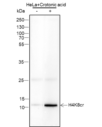
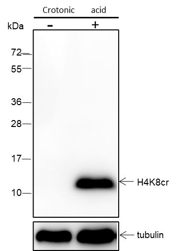
Histones are subject to a variety of enzyme catalyzed modifications, including acetylation, methylation, phosphorylation, ubiquitylation, etc. Crotonylation of lysine is a newly identified reversible modification controlling chromosome structure and gene transcription. The reversible lysine crotonylation has been well demonstrated in eukaryotic histones from worm to human. The unique structure and genomic localization of histone lysine crotonylation suggest that it is mechanistically and functionally different from histone lysine acetylation. Specifically, in both human somatic and mouse male germ cell genomes, histone crotonylation marks either active promoters or potential enhancers. Crotonylation of histone H4 at Lys8 may play a vital role in the epigenetic modulation, including chromatin remodeling and DNA transcriptional regulation.
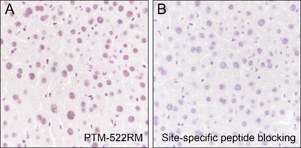
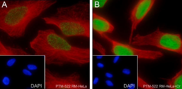
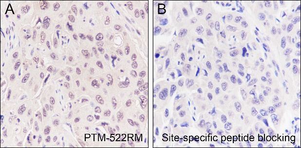
Cellular location

Nucleus

Images
Dot Blot

Blocking buffer: 5% NFDM/TBST
Primary Ab dilution: 1:2000
Primary Ab incubation condition: 2 hours at room temperature
Secondary Ab: Goat Anti-Rabbit IgG H&L pAb (HRP Conjugate)
Immunogen peptide quantity: 4 ng, 16 ng, 64 ng
Exposure time: 60 s
The list of peptides is included in the table below.

WB

Blocking buffer: 5% NFDM/TBST
Primary Ab dilution: 1:2000
Primary Ab incubation condition: 2 hours at room temperature
Secondary Ab: Goat Anti-Rabbit IgG H&L pAb (HRP Conjugate)
Lysate: (-) HeLa, (+) HeLa+Crotonic acid (100 ng/ml, 18hr)
Protein loading quantity: 20 μg
Exposure time: 60 s
Predicted band size: 11 kDa
Observed band size: 11 kDa

IHC-P

Tissue: Human lung squamous carcinoma
Section type: Formalin fixed & Paraffin - embedded section
Retrieval method: High temperature and high pressure
Retrieval buffer: Tris/EDTA buffer, pH 9.0
Primary Ab dilution: 1:500
Primary Ab incubation condition: 1 hour at room temperature
Secondary Ab: Anti-Rabbit and Mouse Polymer HRP (Ready to Use)
Counter stain: Hematoxylin (Blue)
Description: The brown color represents the positive signal observed with PTM-522RM

Tissue: Mouse liver
Section type: Formalin fixed & Paraffin - embedded section
Retrieval method: High temperature and high pressure
Retrieval buffer: Tris/EDTA buffer, pH 9.0
Primary Ab dilution: 1:500
Primary Ab incubation condition: 1 hour at room temperature
Secondary Ab: Anti-Rabbit and Mouse Polymer HRP (Ready to Use)
Counter stain: Hematoxylin (Blue)
Description: The brown color represents the positive signal observed with PTM-522RM

Tissue: Rat kidney
Section type: Formalin fixed & Paraffin - embedded section
Retrieval method: High temperature and high pressure
Retrieval buffer: Tris/EDTA buffer, pH 9.0
Primary Ab dilution: 1:500
Primary Ab incubation condition: 1 hour at room temperature
Secondary Ab: Anti-Rabbit and Mouse Polymer HRP (Ready to Use)
Counter stain: Hematoxylin (Blue)
Description: The brown color represents the positive signal observed with PTM-522RM

ICC/IF

Cell line: (A) HeLa, (B) HeLa+Crotonic acid (100 ng/ml, 18hr)
Fixative: 100% Ice-cold methanol
Permeabilization: 0.1% TritonX-100
Primary Ab dilution: 1:50
Primary incubation condition: 4°C overnight
Secondary Ab: Goat Anti-Rabbit IgG
Nuclear counter stain: DAPI (Blue)
Counter stain: Tubulin (Red)
Description: The green color represents the positive signal observed with PTM-522RM

ChIP

Cell type: HeLa+Crotonic(10mM, 15h)(+)
Cross-linking conditions: No cross-linking
Amount of chromatin per IP: 5×10⁶
cells
Amount of Ab per IP: 6 μg
Beads type and amount per IP: 50 μL of
Protein A MagBeads
Comment:
The ChIP was performed with 6 μg of normal Ra
bbit IgG as a negative control. Real time
quantitative PCR was performed on
immunoprecipitated DNA using primers specific
for the human GAPDH-CDS, RPL30, LDHA-p,
LDHA, Foxo3a-p, Foxo3a-D, RAB20 and
TuBBP10. Data are presented as enrichment of
each sample relative to total amount of input
chromatin at each amplicon.

Research Use

For research use only, not for use in diagnostic procedures.