/ EN
Technical Support
| Contact Us
+1 312-802-6843
Technical Support
Contact Us
+1 312-802-6843
/ EN
Recover password
Professional Email
Verification Code
Send Code
Password
Recombinant Rabbit Monoclonal
Anti-D-Lactyllysine Rabbit mAb
Catalog Number: PTM-1429RM
$ 420

Clone Number: CAPTM-O0804

Host: Rabbit Clonality: Recombinant Monoclonal

Applications: WB ICC/IF IP

Reactivity: Human

Synonyms: Kla, D-la

Product Size
100 μl
Quantity

Shipping: Ambient temperature

Order online or send purchase order to info@ptmbio.com

FAQ Technical Support Protocols

General Information
Isotype IgG
Conjugate Unconjugated
Synonyms Kla, D-la
UniProt ID

/

Immunogen D-lactylated lysine peptides
MW (kDa) Multiple
Specificity Anti-D-Lactyllysine Rabbit mAb detects proteins with D-lactylated lysine residues, with no cross-reactivity to its isomers L-lactyllysine or N-ε-(carboxyethyl)-lysine (Kce). This pan antibody recognizes D-lactylated lysine regardless of the surrounding amino acid sequences.
Product Usage Information
Applications Dilution Recommended Species
WB 1:1000 Human
ICC/IF 1:100 Human
IP 1:50 - 1:100 Human
Properties
Purity Protein A purified
Constituents PBS, Glycerol, BSA
Storage Store at -20°C. Avoid freeze/thaw cycles.
Stability Stable for 12 months from date of receipt/reconstitution.
Target Information

Background

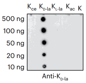
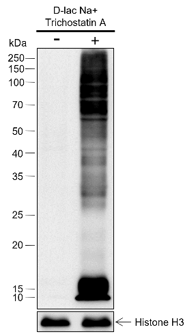
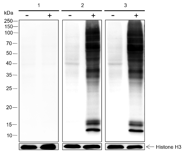
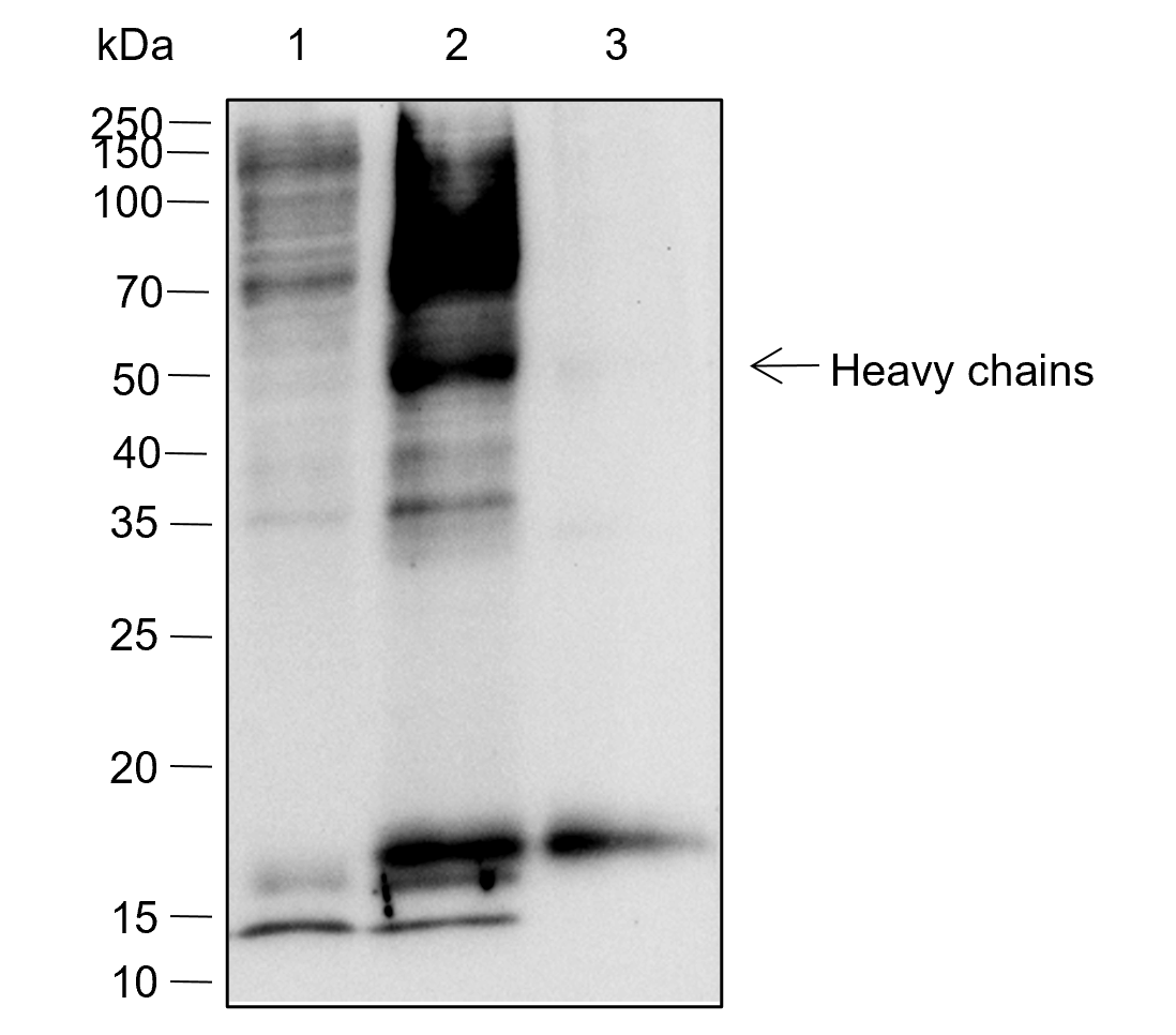
D-lactyllysine (KD-la) refers to the modification of lysine residues in proteins by the addition of D-lactate.dues in proteins. This modification was first reported by Zhang et al. in 2024, with the findings published in Nature Chemical Biology. KD-la forms through a non-enzymatic reaction between proteins and S-D-lactoylglutathione (LGSH), which is a byproduct of the glyoxalase pathway. This pathway involves two key enzymes: glyoxalase 1 (GLO1) and glyoxalase 2 (GLO2). GLO1 conjugates methylglyoxal (MGO), a glycolysis byproduct, with glutathione to form LGSH, which is hydrolyzed by GLO2 to produce D-lactate while regenerating cellular glutathione. In humans, L-lactate is a common metabolic product that plays a key role in energy metabolism, especially during muscle activity. In contrast, D-lactate is not a typical product of human metabolism but is mainly produced by microorganisms such as certain bacteria and yeasts during fermentation. The potential harm of D-lactate mainly lies in its accumulation under specific conditions, such as intestinal microbiota dysbiosis, short bowel syndrome, or certain disease states. Excessive production of D-lactate can lead to D-lactic acidosis, characterized by symptoms of acidosis (low blood bicarbonate levels, decreased blood pH, and hyperuricemia), and in severe cases, neurological symptoms like seizures, ataxia, and altered consciousness.
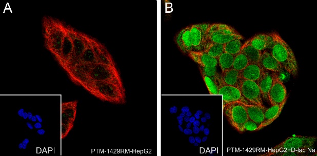
Cellular location

/

Images
Dot Blot

Peptide amount: 4 ng, 20 ng, 50 ng, 200ng
Blocking buffer: 5% NFDM/TBST
Primary Ab dilution: 1:1000
Primary Ab incubation: 2 hours at room temperature
Secondary Ab: Goat Anti-Rabbit IgG H&L pAb (HRP Conjugate)
Exposure time: 60 seconds
The list of peptides used in the experiment is provided below.
Lane 1: D-lactylated peptide library. Lane 2: L-lactylated peptide library.
Lane 3: Carboxyethylated peptide library. Lane 4: Acetylated peptide library.
Lane 5: Butyrylated peptide library. Lane 6: Crotonylated peptide library.
Lane 7: 2-Hydroxyisobutyrylated peptide library. Lane 8: Succinylated peptide library.
Lane 9: Unmodified peptide library.

WB

Lysates: (-): HepG2 cells; (+): HepG2 cells + serum deprivation (0%, 47 hours) add soidum D-lactate (100 mM, 23 hours) add trichostatin A (1 µM, 5 hours)
Protein loading amount: 20 μg
Blocking buffer: 5% NFDM/TBST
Primary Ab dilution: 1:2000
Primary Ab incubation: 2 hours at room temperature
Secondary Ab: Goat Anti-Rabbit IgG H&L pAb (HRP Conjugate)
Exposure time: 60 seconds
Predicted band size: Multiple
Observed band size: Multiple

Lysates: (-): HepG2 cells; (+): HepG2 cells treated with 100 mM soidum D-lactate for 18 hours
Protein loading amount: 20 μg
Blocking buffer: 5% NFDM/TBST
Primary Ab: 1. PTM-1429RM pre-adsorbed with 3 μM synthetic D-lactylated peptides; 2. PTM-1429RM pre-adsorbed with 3 μM synthetic unmodified peptides; 3. PTM-1429RM with no peptide blocking
Primary Ab dilution: 1:1000
Primary Ab incubation: 4°C overnight
Secondary Ab: Goat Anti-Rabbit IgG H&L pAb (HRP Conjugate)
Exposure time: 60 seconds
Predicted band size: Multiple
Observed band size: Multiple

ICC/IF

Samples: (A) HepG2 cells; (B) HepG2 cells treated with 100 mM soidum D-lactate for 18 hours
Fixative: 100% Ice-cold methanol
Permeabilization: 0.1% Triton X-100
Primary Ab dilution: 1:100
Primary Ab incubation: 4°C overnight
Secondary Ab: Goat Anti-Rabbit IgG
Nuclear counter stain: DAPI (blue)
Counter stain: Tubulin (red)
Description: The green color represents the positive signal observed with PTM-1429RM.

IP

IP of HepG2 + soidum D-lactate (100 mM, 18 hours) extracts
Lane 1: 5% Input
Lane 2: IP with PTM-1429RM
Lane 3: IP with Rabbit mAb IgG Isotype Control
IP Ab incubation: 4°C overnight, 1:100 dilution
Blocking buffer: 5% NFDM/TBST
Dilution buffer: 5% NFDM/TBST
WB Primary Ab incubation: PTM-1429RM, 2 hours at room temperature, 1:1000 dilution
Secondary Ab: Anti-Rabbit IgG for IP (HRP Conjugate)
Exposure time: 60 seconds
Predicted band size: Multiple
Observed band size: Multiple

Research Use

For research use only, not for use in diagnostic procedures.