/ EN
Technical Support
| Contact Us
+1 312-802-6843
Technical Support
Contact Us
+1 312-802-6843
/ EN
Recover password
Professional Email
Verification Code
Send Code
Password
Recombinant Rabbit Monoclonal
Anti-Iba1/AIF-1 Rabbit mAb
Catalog Number: PTM-7108
$ 285

Clone Number: JRMR-10822-53B

Host: Rabbit Clonality: Recombinant Monoclonal

Applications: IHC-P mIHC-P

Reactivity: Human

Synonyms: AIF1, G1

Product Size
100 μl
Quantity

Shipping: Ambient temperature

Order online or send purchase order to info@ptmbio.com

FAQ Technical Support Protocols

General Information
Isotype IgG
Conjugate Unconjugated
Synonyms AIF1, G1
UniProt ID

P55008

Immunogen
MW (kDa)
Specificity
Product Usage Information
Applications Dilution Recommended Species
IHC-P 1:500 - 1:1000 Human
Properties
Purity Protein A purified
Constituents PBS, Glycerol, BSA
Storage Store at -20°C. Avoid freeze/thaw cycles.
Stability Stable for 12 months from date of receipt/reconstitution.
Target Information

Background

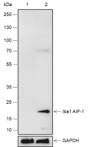
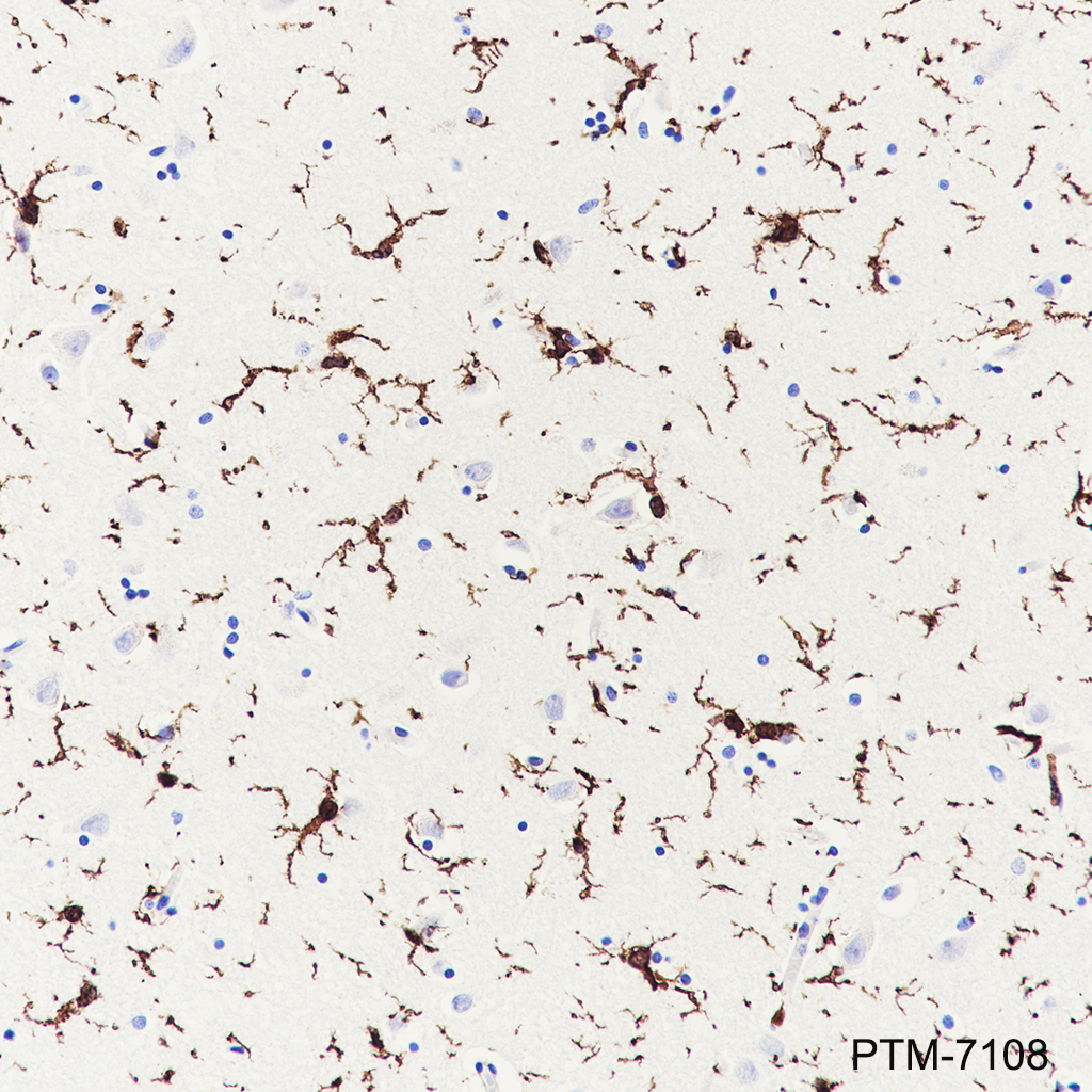
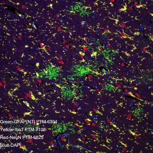
Ionized calcium-binding adaptor molecule 1 (Iba1), also known as allograft inflammatory factor 1 (AIF-1), is an evolutionarily conserved cytoplasmic calcium binding protein containing a central pair of EF-hand calcium binding motifs.Iba1 was originally cloned from activated macrophages in human atherosclerotic allogenic heart grafts undergoing chronic transplant rejection as well as from rat monocytes. Its function is not very well understood, but Iba1 expression is upregulated in response to interferon-gamma and, therefore, could modulate macrophage-dependent immune response. As an F-actin-binding protein, Iba1 may function to remodel the actin cytoskeleton and contribute to morphological changes that correlate with various microglial/macrophage states. Iba1 is also uniquely expressed in cells of monocytic lineage and is, therefore, widely used as a marker for microglia/macrophages in the brain and other tissue.

Cellular location

Cytoplasm, Cytoskeleton

Images
IHC-P

Tissue: Human cerebrum
Section type: Formalin-fixed & paraffin-embedded section
Retrieval method: High temperature and high pressure
Retrieval buffer: Tris/EDTA buffer, pH 9.0
Primary Ab dilution: 1:1000
Primary Ab incubation: 1 hour at room temperature
Secondary Ab: Anti-Rabbit and Mouse Polymer HRP (Ready to Use)
Counter stain: Hematoxylin (blue)
Description: The brown color represents the positive signal observed with PTM-7108

mIHC-P

Tissue: Human cerebrum Section type: Formalin fixed & Paraffin -embedded section Retrieval method: High temperature and high pressure Retrieval buffer: Tris/EDTA buffer, pH 9.0. Heat mediated antigen retrieval (Tris/EDTA buffer, pH 9.0, 20 minutes) was used in between rounds of tyramide signal amplification to remove the antibody from the previous round, to avoid any cross-reactivity. Primary ab dilution: PTM-6104 (1:1000), PTM-7108 (1:1000), PTM-6825 (1:500) Primary Ab incubation: 90 minutes at room temperature Secondary ab: Anti-Rabbit and Mouse Polymer HRP (Ready to use) Nuclear counter stain: DAPI (Blue) Comment: Color red is the positive signal for NeuN (PTM-6825, TSA 570), color green is the positive signal for GFAP(NT) ( PTM-6104, TSA 520), color yellow is the positive signal for lba1 ( PTM-7108, TSA 650). The section was incubated in three rounds of staining; in the order of PTM-6104,PTM-7108,PTM-6825; each using a separate fluorescent tyramide signal amplification system.

Research Use

For research use only, not for use in diagnostic procedures.

Recommended Products
  • View More Images(3)
    $ 285
    Recombinant Rabbit Monoclonal
    Anti-Iba1/AIF-1 Rabbit mAb
    Catalog #
    PTM-6749
    Host
    Rabbit
    Clonality
    Recombinant Monoclonal
    Conjugate
    Unconjugated
    Reactivity
    Human
    Applications:
    WB IHC-P ICC/IF FC IAP IP ChIP mIHC-P