/ EN
Technical Support
| Contact Us
+1 312-802-6843
Technical Support
Contact Us
+1 312-802-6843
/ EN
Recover password
Professional Email
Verification Code
Send Code
Password
Recombinant Rabbit Monoclonal
Anti-L-Lactyl-Histone H3 (Lys23) Rabbit mAb
Catalog Number: PTM-1413RM
$ 435

Clone Number: PA-148(B)-213

Host: Rabbit Clonality: Recombinant Monoclonal

Applications: WB IHC-P ICC/IF ChIP

Reactivity: Human, Mouse, Rat

Synonyms: H3K23la

Product Size
100 μl
Quantity

Shipping: Ambient temperature

Order online or send purchase order to info@ptmbio.com

FAQ Technical Support Protocols

General Information
Isotype IgG
Conjugate Unconjugated
Synonyms H3K23la
UniProt ID

P68431

Immunogen L-lactylated human histone H3 (Lys23) peptide
MW (kDa) 15
Specificity Anti-L-Lactyl-Histone H3 (Lys23) Rabbit mAb detects endogenous levels of histone H3 when it is lactylated at Lys23. This antibody shows minor cross-reactivity to L-lactyl-histone H3 (Lys18) peptide.
Product Usage Information
Applications Dilution Recommended Species
WB 1:2000 - 1:10000 Human, Mouse, Rat
IHC-P 1:200 - 1:1000 Human
ICC/IF 1:100 Human
ChIP 6 μg per 5x10⁶ cells Human
Properties
Purity Protein A purified
Constituents PBS, Glycerol, BSA
Storage Store at -20°C. Avoid freeze/thaw cycles.
Stability Stable for 12 months from date of receipt/reconstitution.
Target Information

Background

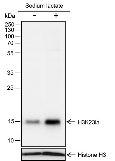
Histones, fundamental proteins involved in chromatin structure and gene regulation, are subject to a wide array of enzyme-catalyzed modifications, including acetylation, methylation, phosphorylation, ubiquitination, and numerous others. Histone L-lactylation, a recently discovered post-translational modification induced by lactate has emerged as a significant addition to this repertoire. The extent and dynamics of this modification are highly reliant on lactate levels within the cellular microenvironment and can be modulated through the introduction of extracellular lactate in cultured cells or the stimulation of intracellular glycolysis. The introduction of lysine L-lactylation is mediated by the acetyltransferase p300, while the removal of lactylation marks from histones has been attributed to Class I histone deacetylases (HDAC 1-3). Histone lactylation has been implicated in various biological processes, including inflammation, fibrosis, differentiation, and cancer progression.

Cellular location

Nucleus

Images
Dot Blot

Peptide amount: 1 ng, 4 ng, 16 ng, 64 ng
Blocking buffer: 5% NFDM/TBST
Primary Ab dilution: 1:2000
Primary Ab incubation condition: 2 hours at room temperature
Secondary Ab: Goat Anti-Rabbit IgG H&L pAb (HRP Conjugate)
Exposure time: 30 seconds
The list of peptides used in the experiment is provided below.
Lane 1: H3K23 lactyl. Lane 2: H3K18 lactyl. Dot 3: H3K23 acetyl.
Dot 4: H3K23 crotonyl. Dot 5: H3K23 butyryl. Dot 6: H3K23 protonyl.
Dot 7: H3K23 unmodified.

WB

Lysates: (-): HeLa cells; (+): HeLa cells treated with 100 mM sodium lactate for 24 hours
Protein loading amount: 20 μg
Blocking buffer: 2.5% BSA/TBST
Primary Ab dilution: 1:10000 (2.5% BSA)
Primary Ab incubation condition: 4°C overnight
Secondary Ab: Goat Anti-Rabbit IgG H&L pAb (HRP Conjugate)
Exposure time: 10 seconds
Predicted band size: 15 kDa
Observed band size: 15 kDa
Important Note: It is recommended to employ 2.5% BSA/TBST for blocking and antibody dilution. Using 5% nonfat milk may result in nonspecific bands.

Lysates: (-): NIH-3T3 cells; (+): NIH-3T3 cells treated with 25 mM sodium lactate for 24 hours
Protein loading amount: 20 μg
Blocking buffer: 2.5% BSA/TBST
Primary Ab dilution: 1:10000 (2.5% BSA)
Primary Ab incubation condition: 4°C overnight
Secondary Ab: Goat Anti-Rabbit IgG H&L pAb (HRP Conjugate)
Exposure time: 10 seconds
Predicted band size: 15 kDa
Observed band size: 15 kDa
Important Note: It is recommended to employ 2.5% BSA/TBST for blocking and antibody dilution. Using 5% nonfat milk may result in nonspecific bands.

Lysates: (-): C6 cells; (+): C6 cells treated with 25 mM sodium lactate for 24 hours
Protein loading amount: 20 μg
Blocking buffer: 2.5% BSA/TBST
Primary Ab dilution: 1:10000 (2.5% BSA)
Primary Ab incubation condition: 4°C overnight
Secondary Ab: Goat Anti-Rabbit IgG H&L pAb (HRP Conjugate)
Exposure time: 10 seconds
Predicted band size: 15 kDa
Observed band size: 15 kDa
Important Note: It is recommended to employ 2.5% BSA/TBST for blocking and antibody dilution. Using 5% nonfat milk may result in nonspecific bands.

IHC-P

Tissue: Human colon
Section type: Formalin fixed & paraffin-embedded section
Retrieval method: High temperature and high pressure
Retrieval buffer: Tris/EDTA buffer, pH 9.0
Primary Ab dilution: 1:1000
Primary Ab incubation condition: 1 hour at room temperature
Secondary Ab: Anti-Rabbit and Mouse Polymer HRP (Ready to Use)
Counter stain: Hematoxylin (blue)
Description: The brown color represents the positive signal observed with PTM-1413RM.

Tissue: Human placenta
Section type: Formalin fixed & paraffin-embedded section
Retrieval method: High temperature and high pressure
Retrieval buffer: Tris/EDTA buffer, pH 9.0
Primary Ab dilution: 1:1000
Primary Ab incubation condition: 1 hour at room temperature
Secondary Ab: Anti-Rabbit and Mouse Polymer HRP (Ready to Use)
Counter stain: Hematoxylin (blue)
Description: The brown color represents the positive signal observed with PTM-1413RM.

ChIP

Sample: HeLa cells + sodium lactate (25 mM, 24 hours)
Cross-linking conditions: No cross-linking
Amount of chromatin per IP: 5×10⁶ cells
Amount of Ab per IP: 6 μg
Beads type and amount per IP: 50 μL of Protein A MagBeads
Description: Chromatin immunoprecipitations were performed with 6 μg of normal rabbit IgG as a negative control. The immunoprecipitated DNA was quantified by real-time PCR using primers specific for the human RPL30, LDHA-promoter, FOXO3a -promoter, FOXO3a-downstream, RAB20, and TUBBP10 regions. The data are presented as enrichment of each sample relative to total amount of input at each amplicon.

Research Use

For research use only, not for use in diagnostic procedures.