/ EN
Technical Support
| Contact Us
+1 312-802-6843
Technical Support
Contact Us
+1 312-802-6843
/ EN
Recover password
Professional Email
Verification Code
Send Code
Password
Recombinant Rabbit Monoclonal
Anti-L-Lactyllysine Rabbit mAb (BSA and Azide Free)
Catalog Number: PTM-1425RM
$ 630

Clone Number: /

Host: Rabbit Clonality: Recombinant Monoclonal

Applications: WB IHC-P ICC/IF FC IP ChIP

Reactivity: All

Synonyms: Kla, L-la

Product Size
100μg
Quantity

Shipping: Ambient temperature

Order online or send purchase order to info@ptmbio.com

FAQ Technical Support Protocols

General Information
Isotype IgG
Conjugate Unconjugated
Synonyms Kla, L-la
UniProt ID

/

Immunogen L-lactylated lysine peptides
MW (kDa) Multiple
Specificity Anti-L-Lactyllysine Rabbit pAb (BSA and Azide Free) detects proteins post-translationally modified by L-lactylation on lysine residues. This pan antibody recognizes L-lactylated lysine independent of its surrounding sequences.
Product Usage Information
Applications Dilution Recommended Species
WB 1:500-1:1000 All
IHC-P 1:200-1:1000 Human, Mouse
ICC/IF 1:50-1:100 Human
FC 1:50-1:100 Human
IP 1:25-1:100 Human
ChIP 6 μg per 5x10⁶ cells Human
Properties
Purity Protein A and immunogen affinity purified
Constituents PBS
Storage Centrifuge the vial at 12,000 x g for 20 seconds before reconstitution. Add 100 μL of sterile deionized water to achieve a final antibody concentration of 1 mg/ml. After reconstitution, the antibody will be in 1X PBS. Aliquot and store at -80°C, and avoid freeze/thaw cycles.
Stability Stable for 12 months from date of receipt/reconstitution.
Target Information

Background

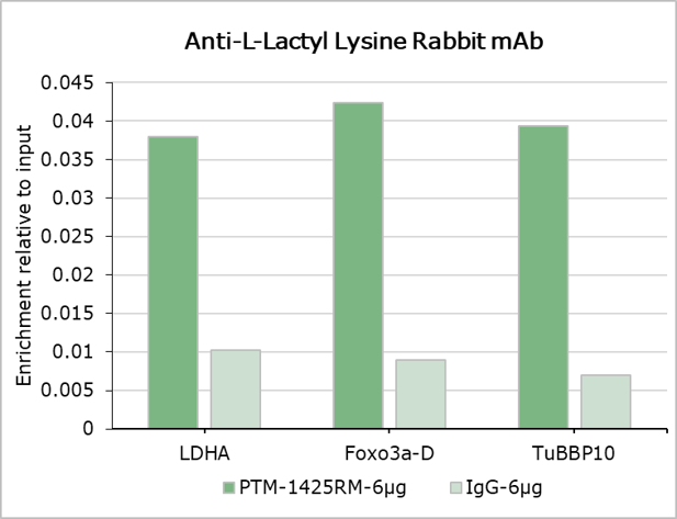
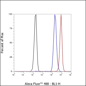
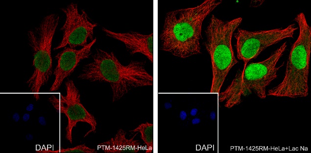
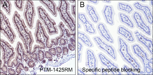
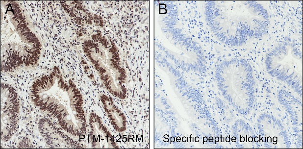
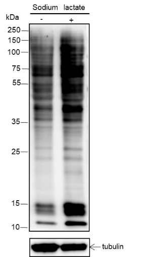
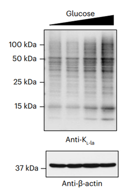
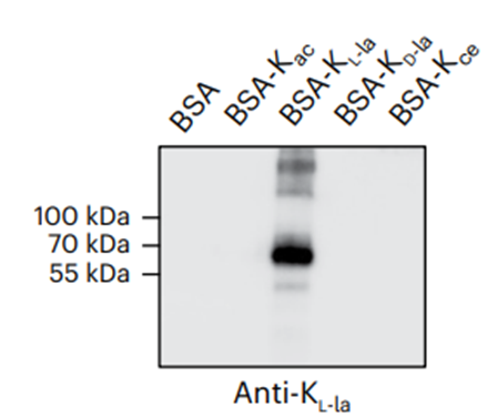
Lactate, previously recognized solely as an energy source and metabolic byproduct, has now emerged as a crucial player in cancer biology, especially within the scope of the Warburg effect and its intricate interplay with various cellular processes, including angiogenesis, hypoxia, polarization of macrophages, and T cell activation. Recent investigations have unveiled an unprecedented function of lactate in the realm of histone modification, specifically through lysine L-lactylation, which exerts regulatory control over gene expression. The extent and dynamics of this modification are highly reliant on lactate levels within the cellular microenvironment and can be modulated through the introduction of extracellular lactate in cultured cells or the stimulation of intracellular glycolysis. The acetyltransferase p300 is responsible for introducing lysine L-lactylation, while Class I histone deacetylases (HDAC 1-3) have been identified as an eraser of the lactylation marks on histones. This BSA-, glycerol-, and azide-free antibody is particularly suitable for applications such as mass cytometry.

Cellular location

/

Images
Dot Blot

Peptide amount: 4 ng, 20 ng, 100 ng
Blocking buffer: 5% NFDM/TBST
Primary Ab dilution: 1:2000
Primary Ab incubation condition: 2 hours at room temperature
Secondary Ab: Goat Anti-Rabbit IgG H&L pAb (HRP Conjugate)
Exposure time: 15 seconds
The list of peptides used in the experiment is provided below.
Lane 1: L-lactylated peptide library. Lane 2: acetylated peptide library.
Lane 3: propionylated peptide library. Lane 4: butyrylated peptide library.
Lane 5: crotonylated peptide library. Lane 6: malonylated peptide library.
Lane 7: 2-hydroxyisobutyrylated peptide library. Lane 8: succinylated peptide library.
Lane 9: unmodified peptide library.

Peptide amount: 10 ng, 20 ng, 50 ng, 100 ng, 500 ng
Blocking buffer: 3% BSA/TBST
Primary Ab dilution: 1:1500
Primary Ab incubation condition: 1 hour at room temperature
Secondary Ab: Goat Anti-Rabbit IgG H&L pAb (HRP Conjugate)
Exposure time: 15 seconds
The list of peptides used in the experiment is provided below.
Kce: carboxyethylated peptide library. KD-la: D-lactylated peptide library.
KL-la: L-lactylated peptide library. Kac: acetylated peptide library.
K: Unmodified peptide library.

Peptide amount: 10 ng, 20 ng, 50 ng, 100 ng, 500 ng
Blocking buffer: 3% BSA/TBST
Primary Ab dilution: 1:1500
Primary Ab incubation condition: 1 hour at room temperature
Secondary Ab: Goat Anti-Rabbit IgG H&L pAb (HRP Conjugate)
Exposure time: 15 seconds
The peptides used in the experiment are synthetic, sequence-specific histone
peptides with the following modifications:
KL-la: L-lactylated peptide library. KD-la: D-lactylated peptide library.
Kce: carboxyethylated peptide library.

WB

Samples: BSA, Acetylated BSA, L-lactylated BSA, D-lactylated BSA, carboxyethylated BSA
Blocking buffer: 3% BSA/TBST
Primary Ab dilution: 1:1500
Primary Ab incubation condition: 1 hour at room temperature
Secondary Ab: Goat Anti-Rabbit IgG H&L pAb (HRP Conjugate)
Exposure time: 15 seconds

Lysates: MCF-7 cells treated with 0.1, 1, 5, and 25 mM glucose
Blocking buffer: 3% BSA/TBST
Primary Ab dilution: 1:1500
Primary Ab incubation condition: 1 hour at room temperature
Secondary Ab: Goat Anti-Rabbit IgG H&L pAb (HRP Conjugate)
Exposure time: 30 seconds
Predicted MW: Multiple
Observed MW: Multiple

Lysates: (-): HeLa cells; (+): HeLa cells treated with 100 mM sodium lactate for 24 hours
Protein loading amount: 20 μg
Blocking buffer: 5% NFDM/TBST
Primary Ab dilution: 1:1000
Primary Ab incubation condition: 2 hours at room temperature
Secondary Ab: Goat Anti-Rabbit IgG H&L pAb (HRP Conjugate)
Exposure time: 30 seconds
Predicted band size: Multiple
Observed band size: Multiple

IHC-P

Tissue: Human colon cancer
Section type: Formalin fixed & paraffin-embedded section
Retrieval method: High temperature and high pressure
Retrieval buffer: Tris/EDTA buffer, pH 9.0
Primary Ab dilution: 1:1000
Primary Ab incubation condition: 1 hour at room temperature
Secondary Ab: Anti-Rabbit and Mouse Polymer HRP (Ready to Use)
Counter stain: Hematoxylin (blue)
Description: The brown color represents the positive signal observed with PTM-1425RM

Tissue: Mouse intestine
Section type: Formalin fixed & paraffin-embedded section
Retrieval method: High temperature and high pressure
Retrieval buffer: Tris/EDTA buffer, pH 9.0
Primary Ab dilution: 1:1000
Primary Ab incubation condition: 1 hour at room temperature
Secondary Ab: Anti-Rabbit and Mouse Polymer HRP (Ready to Use)
Counter stain: Hematoxylin (blue)
Description: The brown color represents the positive signal observed with PTM-1425RM

ICC/IF

Samples: (A) HeLa cells; (B) HeLa cells treated with 100 mM sodium lactate for 24 hours
Fixative: 100% Ice-cold methanol
Permeabilization: 0.1% Triton X-100
Primary Ab dilution: 1:100
Primary Ab incubation condition: 4°C overnight
Secondary Ab: Goat Anti-Rabbit IgG
Nuclear counter stain: DAPI (blue)
Counter stain: Tubulin (red)
Description: The green color represents the positive signal observed with PTM-1425RM.

FC

Sample: HeLa cells
Fixative: 4% Paraformaldehyde
Permeabilization: 0.1% Triton X-100
Primary Ab dilution: 1:100
Secondary Ab: Goat Anti-Rabbit IgG
Unlabeled control: The cell without incubation with primary antibody and secondary antibody (black line).
Isotype control: Rabbit mAb IgG Isotype Control (blue line).
Description: The red line represents the positive signal observed with PTM-1425RM.

IP

IP of HeLa + sodium lactate (100 mM, 24 hours) extracts
Lane 1: 5% Input
Lane 2: IP with PTM-1425RM
Lane 3: IP with Rabbit mAb IgG Isotype Control
IP Ab incubation condition: 4°C overnight, 1:100 dilution
Blocking buffer: 5% NFDM/TBST
Dilution buffer: 5% NFDM/TBST
WB primary Ab incubation condition: PTM-1425RM, 2 hours at room temperature, 1:1000 dilution
Secondary Ab: Anti-Rabbit IgG for IP (HRP Conjugate)
Exposure time: 60 seconds
Predicted band size: Multiple
Observed band size: Multiple

ChIP

Sample: MCF-7 cells
Cross-linking conditions: no cross-linking
Amount of chromatin per IP: 5×10⁶ cells
Amount of Ab per IP: 6 μg
Beads type and amount per IP: 50 μL of Protein A/G MagBeads
Description: Chromatin immunoprecipitations were performed with 6 μg of normal rabbit IgG as a negative control. The immunoprecipitated DNA was quantified by real-time PCR using primers specific for the human LDHA CDS, FOXO3a downstream, and TUBBP10 regions. The data are presented as enrichment of each sample relative to total amount of input at each amplicon.

Research Use

For research use only, not for use in diagnostic procedures.