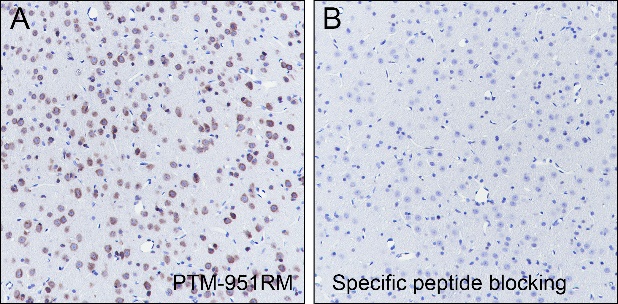
/ EN
Technical Support
| Contact Us
+1 312-802-6843
Technical Support
Contact Us
+1 312-802-6843
/ EN
Recover password
Professional Email
Verification Code
Send Code
Password
Recombinant Rabbit Monoclonal
Anti-O-Linked N-Acetylglucosamine Rabbit mAb
Catalog Number: PTM-951RM
$ 340

Clone Number: 17H2L2

Host: Rabbit Clonality: Recombinant Monoclonal

Applications: WB IHC-P ICC/IF

Reactivity: Human, Mouse, Rat

Synonyms: O-GlcNAc

Product Size
100 μl
Quantity

Shipping: Ambient temperature

Order online or send purchase order to info@ptmbio.com

FAQ Technical Support Protocols

General Information
Isotype IgG
Conjugate Unconjugated
Synonyms O-GlcNAc
UniProt ID

/

Immunogen
MW (kDa) Multiple
Specificity Anti-O-Linked N-Acetylglucosamine Rabbit mAb detects proteins with O-Linked N-Acetylglucosamine residues. This pan antibody recognizes O-Linked N-Acetylglucosamine independent of its surrounding sequences.
Product Usage Information
Applications Dilution Recommended Species
WB 1:500 - 1:1000 All
ICC/IF 1:50 Human
IHC-P 1:200-1:1000 Mouse、Rat
Properties
Purity Protein A purified
Constituents PBS, Glycerol, BSA
Storage Store at -20°C. Avoid freeze/thaw cycles.
Stability Stable for 12 months from date of receipt/reconstitution.
Target Information

Background

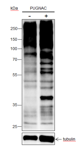
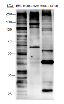
O-Linked β-N-acetylglucosamine modification (GlcNAcylation) is a glycosylation in which the monosaccharide β-N-acetylglucosamine (GlcNAc) attaches to serine/threonine residues via an O-linked glycosidic bond and is found mostly within the cytoplasm or nucleoplasm. GlcNAcylation regulates several important cellular processes, such as signal transduction, protein expression, degradation, embryonic stem cell pluripotency and trafficking. This post-translational modification is regulated by OGT and O-GlcNAcase (β-N-acetylglucosaminidase) enzymes, whereas OGT catalyzes the attachment and O-GlcNAcase catalyzes the removal of O-GlcNAc to proteins. O-GlcNAc modification has been observed on histones: H2A at T101, H2B at S36 and S112, H3 at S10 and T32, and H4 at S47.
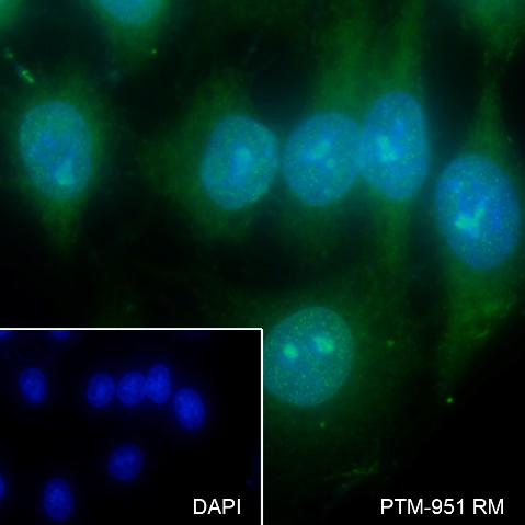
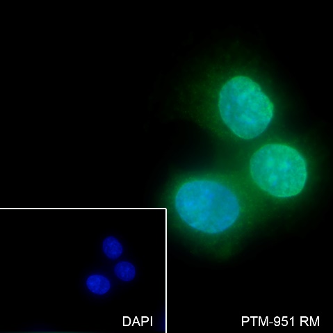
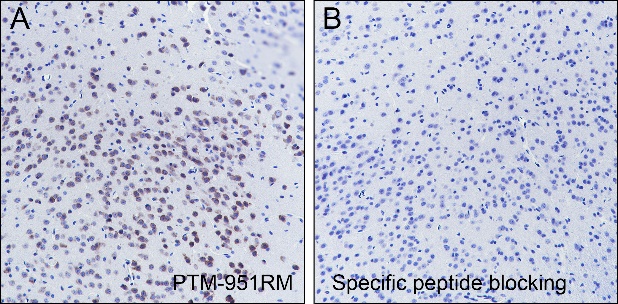
Cellular location

/

Images
Dot Blot

Blocking buffer: 5% NFDM/TBST
Primary Ab dilution: 1:2000
Primary Ab incubation condition: 2 hours at room temperature
Secondary Ab: Goat Anti-Rabbit IgG H&L pAb (HRP Conjugate)
Immunogen peptide quantity: 4 ng, 20 ng, 100 ng
Exposure time: 60 s
The list of peptides is included in the table below.

WB

Blocking buffer: 5% NFDM/TBST
Primary Ab dilution: 1:1000
Primary Ab incubation condition: 2 hours at room temperature
Secondary Ab: Goat Anti-Rabbit IgG H&L pAb (HRP Conjugate)
Lysate: (-) HeLa, (+) HeLa+PUGNAC (100μM, 24hr)
Protein loading quantity: 20 μg
Exposure time: 60 s
Predicted band size: Multiple kDa
Observed band size: Multiple kDa

Blocking buffer: 5% NFDM/TBST
Primary Ab dilution: 1:1000
Primary Ab incubation condition: 2 hours at room temperature
Secondary Ab: Goat Anti-Rabbit IgG H&L pAb (HRP Conjugate)
Lysate: BRL, Mouse liver, Mouse colon
Protein loading quantity: 20 μg
Exposure time: 60 s
Predicted band size: Multiple kDa
Observed band size: Multiple kDa

ICC/IF

Cell line: HepG2
Fixative: 100% Ice-cold methanol
Permeabilization: 0.1% TritonX-100
Primary Ab dilution: 1:50
Primary incubation condition: 4°C overnight
Secondary Ab: Goat Anti-Rabbit IgG
Nuclear counter stain: DAPI (Blue)
Description: The green color represents the positive signal observed with PTM-951RM

Cell line: MCF-7
Fixative: 100% Ice-cold methanol
Permeabilization: 0.1% TritonX-100
Primary Ab dilution: 1:50
Primary incubation condition: 4°C overnight
Secondary Ab: Goat Anti-Rabbit IgG
Nuclear counter stain: DAPI (Blue)
Description: The green color represents the positive signal observed with PTM-951RM

IHC-P

Tissue: Mouse cerebrum
Section type: Formalin-fixed & Paraffinembedded section
Retrieval method: High temperature and high
pressure
Retrieval buffer: Tris/EDTA buffer, pH 9.0
Primary Ab dilution: 1:1000
Primary Ab incubation condition: 1 hour at
room temperature
Secondary Ab: Anti-Rabbit and Mouse Polymer
HRP (Ready to use)
Counter stain: Hematoxylin (Blue)
Comment: Color brown is the positive signal for
PTM-951RM

Tissue: Rat cerebrum
Section type: Formalin-fixed & Paraffinembedded section
Retrieval method: High temperature and high
pressure
Retrieval buffer: Tris/EDTA buffer, pH 9.0
Primary Ab dilution: 1:1000
Primary Ab incubation condition: 1 hour at
room temperature
Secondary Ab: Anti-Rabbit and Mouse Polymer
HRP (Ready to use)
Counter stain: Hematoxylin (Blue)
Comment: Color brown is the positive signal for
PTM-951RM

Research Use

For research use only, not for use in diagnostic procedures.

Recommended Products
  • View More Images(3)
    $ 350
    Anti-O-Linked N-Acetylglucosamine Mouse mAb
    Catalog #
    PTM-952
    Host
    Mouse
    Clonality
    Monoclonal
    Conjugate
    Unconjugated
    Reactivity
    Human, Mouse, Rat
    Applications:
    WB IHC-P ICC/IF FC IAP IP ChIP mIHC-P