/ EN
Technical Support
| Contact Us
+1 312-802-6843
Technical Support
Contact Us
+1 312-802-6843
/ EN
Recover password
Professional Email
Verification Code
Send Code
Password
Recombinant Rabbit Monoclonal
Anti-PD-L1 Rabbit mAb
Catalog Number: PTM-6103
$ 280

Clone Number: 10479

Host: Rabbit Clonality: Monoclonal

Applications: WB IHC-P

Reactivity: Human

Synonyms: CD274, B7H1, PDCD1L1, PDCD1LG1

Product Size
100μl
Quantity

Shipping: Ambient temperature

Order online or send purchase order to info@ptmbio.com

FAQ Technical Support Protocols

General Information
Isotype IgG/kappa
Conjugate Unconjugated
Synonyms CD274, B7H1, PDCD1L1, PDCD1LG1
UniProt ID

Q9NZQ7

Immunogen
MW (kDa)
Specificity
Product Usage Information
Applications Dilution Recommended Species
WB 1:500-1:1000 Human
IHC-P 1:50-1:100 Human
Properties
Purity Protein G purified
Constituents PBS, Glycerol, BSA
Storage Store at -20°C. Avoid freeze/thaw cycles.
Stability Stable for 12 months from date of receipt/reconstitution.
Target Information

Background

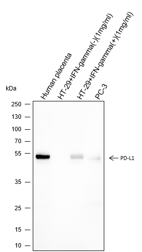
Programmed death-ligand 1 (PD-L1) also known as cluster of differentiation 274 (CD274) or B7 homolog 1 (B7-H1) is a protein that in humans is encoded by the CD274 gene. Programmed death-ligand 1 (PD-L1) is a type 1 transmembrane protein that has been speculated to play a major role in suppressing the adaptive arm of immune system during particular events such as pregnancy, tissue allografts, autoimmune disease and other disease states such as hepatitis. The binding of PD-L1 to the inhibitory checkpoint molecule PD-1 transmits an inhibitory signal based on interaction with phosphatases (SHP-1 or SHP-2) via Immunoreceptor Tyrosine-Based Switch Motif (ITSM).This reduces the proliferation of antigen-specific T-cells in lymph nodes, while simultaneously reducing apoptosis in regulatory T cells (anti-inflammatory, suppressive T cells) - further mediated by a lower regulation of the gene Bcl-2.
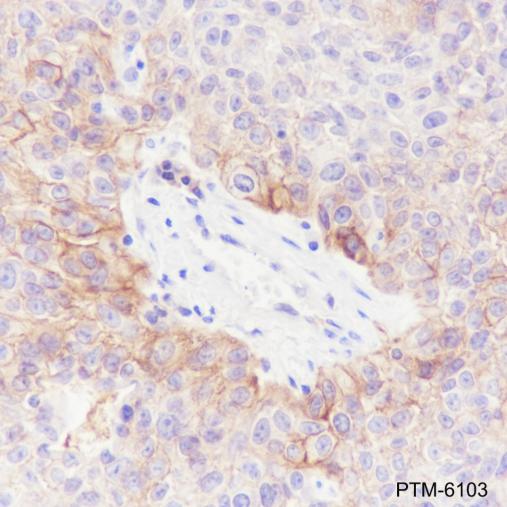
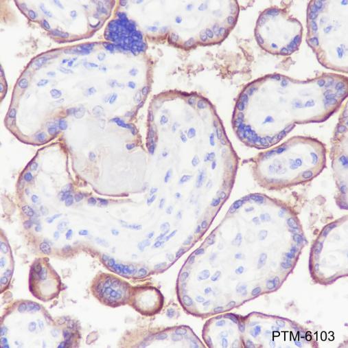
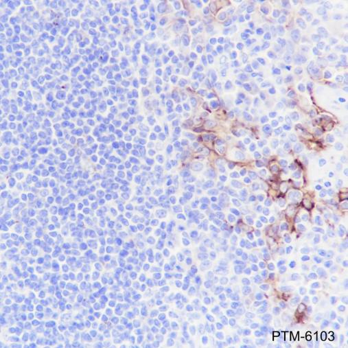
Cellular location

Cell membrane, Endosome

Images
WB

Blocking buffer: 5% NFDM/TBST
Primary ab dilution: 1:1000
Primary ab incubation condition: 2 hours at room temperature
Secondary ab: Goat Anti-Rabbit IgG H&L (HRP)
Lysate: Human placenta, HT-29+IFNgamma(-)(1mg/ml), HT-29+IFNgamma(+)(1mg/ml), PC-3
Protein loading quantity: 20 μg
Exposure time: 60 s
Predicted MW: 50 kDa
Observed MW: 50 kDa

IHC-P

Tissue: Human lung squamous carcinoma
Section type: Formalin fixed & Paraffin -embedded section
Retrieval method: High temperature and high pressure
Retrieval buffer: Tris/EDTA buffer, pH 9.0
Primary ab dilution: 1:100
Primary ab incubation condition: 1 hour at room temperature
Secondary ab: Anti-Rabbit and Mouse Polymer HRP (Ready to use)
Counter stain: Hematoxylin (Blue)
Comment: Color brown is the positive signal for PTM-6103

Tissue: Human placenta
Section type: Formalin fixed & Paraffin -embedded section
Retrieval method: High temperature and high pressure
Retrieval buffer: Tris/EDTA buffer, pH 9.0
Primary ab dilution: 1:100
Primary ab incubation condition: 1 hour at room temperature
Secondary ab: Anti-Rabbit and Mouse Polymer HRP (Ready to use)
Counter stain: Hematoxylin (Blue)
Comment: Color brown is the positive signal for PTM-6103

Tissue: Human tonsil
Section type: Formalin fixed & Paraffin -embedded section
Retrieval method: High temperature and high pressure
Retrieval buffer: Tris/EDTA buffer, pH 9.0
Primary ab dilution: 1:100
Primary ab incubation condition: 1 hour at room temperature
Secondary ab: Anti-Rabbit and Mouse Polymer HRP (Ready to use)
Counter stain: Hematoxylin (Blue)
Comment: Color brown is the positive signal for PTM-6103

Research Use

For research use only, not for use in diagnostic procedures.