/ EN
Technical Support
| Contact Us
+1 312-802-6843
Technical Support
Contact Us
+1 312-802-6843
/ EN
Recover password
Professional Email
Verification Code
Send Code
Password
Recombinant Rabbit Monoclonal
Anti-Phospho-Threonine Rabbit mAb
Catalog Number: PTM-705RM
$ 290

Clone Number: PA-031-10

Host: Rabbit Clonality: Recombinant Monoclonal

Applications: WB ICC/IF

Reactivity: All

Synonyms: pThr

Product Size
100 μl
Quantity

Shipping: Ambient temperature

Order online or send purchase order to info@ptmbio.com

FAQ Technical Support Protocols

General Information
Isotype IgG
Conjugate Unconjugated
Synonyms pThr
UniProt ID

/

Immunogen
MW (kDa) Multiple
Specificity Anti-Phospho-Threonine Rabbit mAb detects proteins with Phospho-Threoninated lysine residues. This pan antibody recognizes Phospho-Threoninated lysine independent of its surrounding sequences.
Product Usage Information
Applications Dilution Recommended Species
WB 1:500 - 1:1000 All
ICC/IF 1:50 Huma
Properties
Purity Protein A purified
Constituents PBS, Glycerol, BSA
Storage Store at -20°C. Avoid freeze/thaw cycles.
Stability Stable for 12 months from date of receipt/reconstitution.
Target Information

Background

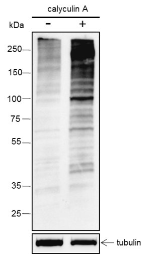
Protein phosphorylation is a common post-translational modification that can occur on many types of amino acids, such as tyrosine, serine, and threonine. The phosphorylation of threonine residues is related to many growth factors and oncogene protein kinases and plays an important role in the signal transduction of protein activation, cell proliferation and differentiation. The study of the transformation mechanism of oncogenes and growth factors in the mitogenic process depends on the identification of their substrates and subsequent determination of how phosphorylation affects their properties. The antibody that specifically recognizes amino acid phosphorylation can quickly identify, quantify, and immunoaffinityly separate cellular phosphorylated proteins, and can be used as an analysis and preparation tool, which is of inestimable value to the above-mentioned research.
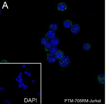
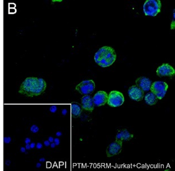
Cellular location

/

Images
Dot Blot

Blocking buffer: 5% NFDM/TBST
Primary Ab dilution: 1:2000
Primary Ab incubation condition: 2 hours at room temperature
Secondary Ab: Goat Anti-Rabbit IgG H&L pAb (HRP Conjugate)
Immunogen peptide quantity: 20 ng, 100 ng
Exposure time: 60 s
The list of peptides is included in the table below.

WB

Blocking buffer: 5% NFDM/TBST
Primary Ab dilution: 1:1000
Primary Ab incubation condition: 2 hours at room temperature
Secondary Ab: Goat Anti-Rabbit IgG H&L pAb (HRP Conjugate)
Lysate:(-): Jurkat, (+): Jurkat+ calyculin A (100nM, 30min)
Protein loading quantity: 20 μg
Exposure time: 60 s
Predicted band size: Multiple
Observed band size: Multiple

Blocking buffer: 5% NFDM/TBST
Primary Ab dilution: 1:1000
Primary Ab incubation condition: 2 hours at room temperature
Secondary Ab: Goat Anti-Rabbit IgG H&L pAb (HRP Conjugate)
Lysate: (-): HeLa, (+): HeLa+ calyculin A (100nM, 15min)
Protein loading quantity: 20 μg
Exposure time: 60 s
Predicted band size: Multiple
Observed band size: Multiple

ICC/IF

Cell line: Jurkat
Fixative: 100% Ice-cold methanol
Permeabilization: 0.1% TritonX-100
Primary ab dilution: 1:50
Primary incubation condition: 4℃ overnight
Secondary ab: Goat Anti-Rabbit IgG
Nuclear counter stain: DAPI (Blue)
Counter stain: Tubulin (Red)
Comment: Color green is the positive signal for
PTM-705RM

Cell line: Jurkat+calyculin A ( 100nM, 30min)
Fixative: 100% Ice-cold methanol
Permeabilization: 0.1% TritonX-100
Primary ab dilution: 1:50
Primary incubation condition: 4℃ overnight
Secondary ab: Goat Anti-Rabbit IgG
Nuclear counter stain: DAPI (Blue)
Counter stain: Tubulin (Red)
Comment: Color green is the positive signal for
PTM-705RM

Research Use

For research use only, not for use in diagnostic procedures.

References (1)
  • p38α in the preoptic area inhibits brown adipose tissue thermogenesis
    Year 2022
    Journal Obesity
    Authors Jing Wang, et al.
    Applications Unspecified
    Reactivity